

PART 7: Oncology and Hematology
SECTION 3 Disorders of Hemostasis
116 Coagulation Disorders
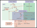 | Figure 116-1 Coagulation cascade and laboratory assessment of clotting factor deficiency by activated partial prothrombin time (aPTT), prothrombin time (PT), and thrombin time (TT). |
view large |
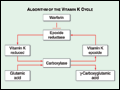 | Figure 116-2 The vitamin K cycle. Vitamin K is a cofactor for the formation of γ-carboxyglutamic acid residues on coagulation proteins. Vitamin K–dependent γ-glutamylcarboxylase, the enzyme that catalyzes the vitamin K epoxide reductase, regenerates reduced vitamin K. Warfarin blocks the action of the reductase... |
view large |
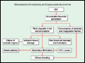 | Figure 116-3 The pathophysiology of disseminated intravascular coagulation (DIC). Interactions between coagulation and fibrinolytic pathways result in bleeding and thrombosis in the microcirculation in patients with DIC. |
view large |
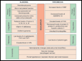 | Figure 116-4 Balance of hemostasis in liver disease. TAFI, thrombin-activated fibriolytic inhibitor; t-PA, tissue plasminogen activator; VWF, von Willebrand factor. |
view large |