

PART 7: Oncology and Hematology
SECTION 3 Disorders of Hemostasis
115 Disorders of Platelets and Vessel Wall
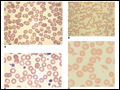 | Figure 115-1 Photomicrographs of peripheral blood smears: A. Normal peripheral blood. B. Platelet clumping in pseudothrombocytopenia. C. Abnormal large platelet in autosomal dominant macrothrombocytopenia. D. Schistocytes and decreased platelets in microangiopathic hemolytic... |
view large |
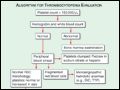 | Figure 115-2 Algorithm for evaluating the thrombocytopenic patient. |
view large |
 | Figure 115-3 Time course of heparin-induced thrombocytopenia (HIT) development after heparin exposure. The timing of development after heparin exposure is a critical factor in determining the likelihood of HIT in a patient. HIT occurs early after heparin exposure in the presence of preexisting heparin/platelet factor 4 (PF4)... |
view large |
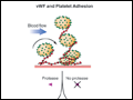 | Figure 115-4 Pathogenesis of thrombotic thrombocytopenic purpura (TTP). Normally the ultra-high-molecular-weight multimers of von Willebrand factor (VWF) produced by the endothelial cells are processed into smaller multimers by a plasma metalloproteinase called ADAMTS13. In TTP the activity of the protease is inhibited, and the... |
view large |
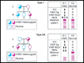 | Figure 115-5 Pattern of inheritance and laboratory findings in von Willebrand disease. The assays of platelet function include a coagulation assay of Factor VIII bound and carried by von Willebrand factor (VWF), abbreviated as VIII; immunoassay of total VWF protein (VWF:Ag); bioassay of the ability of patient plasma to support... |
view large |