

PART 7: Oncology and Hematology
SECTION 2 Hematopoietic Disorders
111 Plasma Cell Disorders
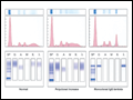 | Figure 111-1 Representative patterns of serum electrophoresis and immunofixation. The upper panels represent agarose gel, middle panels are the densitometric tracing of the gel, and lower panels are immunofixation patterns. Panel on the left illustrates the normal pattern of serum protein on electrophoresis. Since there are many different... |
view large |
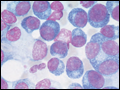 | Figure 111-2 Multiple myeloma (marrow). The cells bear characteristic morphologic features of plasma cells, round or oval cells with an eccentric nucleus composed of coarsely clumped chromatin, a densely basophilic cytoplasm, and a perinuclear clear zone containing the Golgi apparatus. Binucleate and multinucleate malignant plasma cells can be... |
view large |
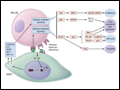 | Figure 111-3 Pathogenesis of multiple myeloma. Multiple myeloma cells interact with bone marrow stromal cells and extracellular matrix proteins via adhesion molecules, triggering adhesion-mediated signaling as well as cytokine production. This triggers cytokine-mediated signaling that provides growth, survival, and antiapoptotic effects as... |
view large |
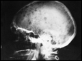 | Figure 111-4 Bony lesions in multiple myeloma. The skull demonstrates the typical "punched out" lesions characteristic of multiple myeloma. The lesion represents a purely osteolytic lesion with little or no osteoblastic activity. (Courtesy of Dr. Geraldine Schechter; with permission.) |
view large |