

PART 7: Oncology and Hematology
SECTION 1 Neoplastic Disorders
101 Paraneoplastic Neurologic Syndromes
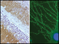 | Figure 101-1 Antibodies to NR1/NR2 subunits of the NMDA receptor in a patient with paraneoplastic encephalitis and ovarian teratoma. Panel A is a section of dentate gyrus of rat hippocampus immunolabeled (brown staining) with the patient's antibodies. The reactivity predominates in the molecular layer, which... |
view large |
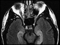 | Figure 101-2 Fluid-attenuated inversion recovery sequence MRI of a patient with limbic encephalitis and LGI1 antibodies. Note the abnormal hyperintensity involving the medial aspect of the temporal lobes. |
view large |
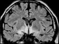 | Figure 101-3 MRI and tumor of a patient with anti-Ma2-associated encephalitis. Panels A and B are fluid-attenuated inversion recovery MRI sequences showing abnormal hyperintensities in the medial temporal lobes, hypothalamus, and upper brainstem. |
view large |