

PART 7: Oncology and Hematology
SECTION 1 Neoplastic Disorders
84 Cancer Cell Biology and Angiogenesis
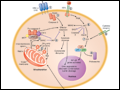
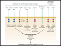
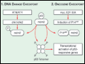 | Figure 84-1 Induction of p53 by the DNA damage and oncogene checkpoints. In response to noxious stimuli, p53 and mdm2 are phosphor-ylated by the ataxia-telangiectasia mutated (ATM) and related ATR serine/threonine kinases, as well as the immediate downstream checkpoint kinases, Chk1 and Chk2. This causes dissociation of p53 from mdm2,... |
view large |
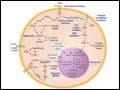 | Figure 84-2 Therapeutic targeting of signal transduction pathways in cancer cells. Three major signal transduction pathways are activated by receptor tyrosine kinases (RTK). 1. The protooncogene Ras is activated by the Grb2/mSOS guanine nucleotide exchange factor, which induces an association with Raf and... |
view large |
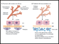
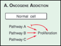 | Figure 84-3 Oncogene addiction and synthetic lethality: keys to discovery of new anticancer drugs. Panel A. Normal cells receive environmental signals that activate signaling pathways (pathways A, B, and C) that together promote G1 to S phase transition and passage through the... |
view large |
 | Figure 84-4 Epigenetic regulation of gene expression in cancer cells. Tumor-suppressor genes are often epigenetically silenced in cancer cells. In the upper portion, a CpG island within the promoter and enhancer regions of the gene has been methylated, resulting in the recruitment of methyl-cytosine binding proteins (MeCP) and complexes... |
view large |
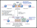
 | Figure 84-5 Therapeutic strategies to overcome aberrant survival pathways in cancer cells.1. The extrinsic pathway of apoptosis can be selectively induced in cancer cells by TRAIL (the ligand for death receptors 4 and 5) or by agonistic monoclonal antibodies. 2. Inhibition of... |
view large |
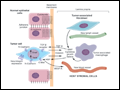 | Figure 84-6 Oncogene signaling pathways are activated during tumor progression and promote metastatic potential. This figure shows a cancer cell that has undergone epithelial to mesenchymal transition (EMT) under the influence of several environmental signals. Critical components include activated transforming growth factor beta... |
view large |
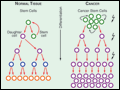 | Figure 84-7 Cancer stem cells play a critical role in the initiation, progression, and resistance to therapy of malignant neoplasms. In normal tissues (left), homeostasis is maintained by asymmetric division of stem cells leading to one progeny cell that will differentiate and one cell that will maintain the stem cell pool. This occurs... |
view large |
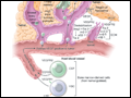 | Figure 84-8 Tumor angiogenesis is a complex process involving many different cell types that must proliferate, migrate, invade, and differentiate in response to signals from the tumor microenvironment. Endothelial cells (ECs) sprout from host vessels in response to VEGF, bFGF, Ang2, and other proangiogenic stimuli. Sprouting is stimulated... |
view large |
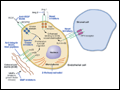
 | Figure 84-9 Critical molecular determinants of endothelial cell biology. Angiogenic endothelium expresses a number of receptors not found on resting endothelium. These include receptor tyrosine kinases (RTKs) and integrins that bind to the extracellular matrix and mediate endothelial cell (EC) adhesion, migration, and invasion. ECs also... |
view large |
 | Figure 84-10 Normalization of tumor blood vessels due to inhibition of VEGF signaling.A. Blood vessels in normal tissues exhibit a regular hierarchical branching pattern that delivers blood to tissues in a spatially and temporally efficient manner to meet the metabolic needs of the... |
view large |
 | Figure 84-11 Knowledge of the molecular events governing tumor angiogenesis has led to a number of therapeutic strategies to block tumor blood vessel formation. The successful therapeutic targeting of VEGF is described in the text. Other endothelial cell–specific receptor tyrosine kinase pathways (e.g., angiopoietin/Tie2 and... |
view large |