

PART 3: Genes, the Environment, and Disease
62 Chromosome Disorders
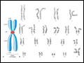 | Figure 62-1 A. An idealized human chromosome, showing the centromere (cen), long (q) and short (p) arms, and telomeres (tel). B. A G-banded human karyotype from a normal (46,XX) female. |
view large |
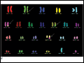 | Figure 62-2 Examples of different applications of fluorescence in situ hybridization (FISH) to human metaphase and interphase preparations. A, B. Aneuploidy detection: Interphase FISH using chromosome 13 (green) and chromosome 21 (red) unique sequence probes on interphase... |
view large |
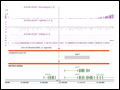 | Figure 62-3 Array analysis to diagnose human chromosome abnormalities. Detection of a 533-kb deletion in 17q21.31 that includes the MAPT gene that is responsible for a newly described microdeletion syndrome. The SNP array illustrates the deletion by changes in the log2 ratio, allele difference, smooth signal, and copy number... |
view large |
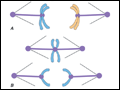 | Figure 62-4 Chromosome segregation in meiosis. A. In meiosis I, each of the 23 pairs of chromosomes finds its "partner," or homologue, and exchanges genetic material (recombines) with it. At metaphase, each homologous pair aligns on the equatorial plate; at anaphase, each member of the... |
view large |
 | Figure 62-5 Use of DNA technology to determine the origin of chromosome abnormalities. A. Analysis of a chromosome 21–specific DNA polymorphism demonstrates that the trisomic individual received two chromosomes 21 from his mother and one from his father; thus, the extra chromosome 21... |
view large |
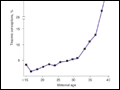 | Figure 62-6 Estimated maternal age–adjusted rates of trisomy among all clinically recognized pregnancies (e.g., spontaneous abortions, stillbirths, and livebirths). Among women in their forties, more than 25% of all pregnancies are estimated to involve a trisomic conception; the vast majority of these spontaneously abort,... |
view large |