

PART 3: Genes, the Environment, and Disease
61 Principles of Human Genetics
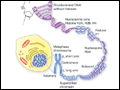 | Figure 61-1 Structure of chromatin and chromosomes. Chromatin is composed of double-strand DNA that is wrapped around histone and non–histone proteins forming nucleosomes. The nucleosomes are further organized into solenoid structures. Chromosomes assume their characteristic structure, with short (p) and long (q) arms at the metaphase stage of the cell... |
view large |
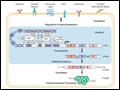 | Figure 61-2 Flow of genetic information. Multiple extracellular signals activate intracellular signal cascades that result in altered regulation of gene expression through the interaction of transcription factors with regulatory regions of genes. RNA polymerase transcribes DNA into RNA that is processed to mRNA by excision of... |
view large |
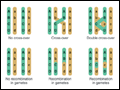 | Figure 61-3 Crossing-over and genetic recombination. During chiasma formation, either of the two sister chromatids on one chromosome pairs with one of the chromatids of the homologous chromosome. Genetic recombination occurs through crossing-over and results in recombinant and nonrecombinant chromosome segments in the gametes.... |
view large |
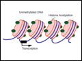 | Figure 61-4 Epigenetic modifications of DNA and histones. Methylation of cytosine residues is associated with gene silencing. Methylation of certain genomic regions is inherited (imprinting) and is involved in the silencing of one of the two X chromosomes in females (X-inactivation). Alterations in methylation can also be acquired... |
view large |
 | Figure 61-5 Point mutations causing β-thalassemia as example of allelic heterogeneity. The β-globin gene is located in the globin gene cluster. Point mutations can be located in the promoter, the CAP site, the 5′-untranslated region, the initiation codon, each of the three... |
view large |
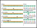 | Figure 61-6 A. Examples of mutations. The coding strand is shown with the encoded amino acid sequence. B. Chromatograms of sequence analyses after amplification of genomic DNA by polymerase chain reaction. |
view large |
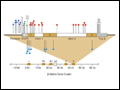
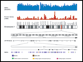 | Figure 61-7 Chromosome 7 is shown with the density of single nucleotide polymorphisms (SNPs) and genes above. A 200-kb region in 7q31.2 containing the CFTR gene is shown below. The CFTR gene contains 27 exons. More than 1790 mutations in this gene have... |
view large |
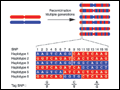 | Figure 61-8 The origin of haplotypes is due to repeated recombination events occurring in multiple generations. Over time, this leads to distinct haplotypes. These haplotype blocks can often be characterized by genotyping selected Tag single-nucleotide polymorphisms, an approach that now facilitates performing genome-wide association studies... |
view large |
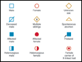 | Figure 61-9 Standard pedigree symbols. |
view large |
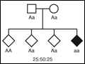 | Figure 61-10 Segregation of alleles. Segregation of genotypes in the offspring of parents with one dominant (A) and one recessive (a) allele. The distribution of the parental alleles to their offspring depends on the combination present in the parents. Filled symbols represent affected individuals. |
view large |
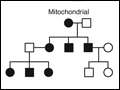 | Figure 61-11 Dominant, recessive, X-linked, and mitochondrial (matrilinear) inheritance. |
view large |
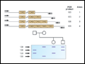 | Figure 61-12 CAG repeat length and linkage analysis in multiple endocrine neoplasia (MEN) type 1. Upper panel. Detection of different alleles using polymorphic microsatellite markers. The example depicts a CAG trinucleotide repeat. PCR with primers flanking the polymorphic region results in... |
view large |
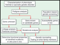 | Figure 61-13 Approach to genetic disease. |
view large |