

PART 2: Cardinal Manifestations and Presentation of Diseases
SECTION 10 Hematologic Alterations
60 Disorders of Granulocytes and Monocytes
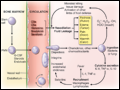 | Figure 60-1 Schematic events in neutrophil production, recruitment, and inflammation. The four cardinal signs of inflammation (rubor, tumor, calor, dolor) are indicated, as are the interactions of neutrophils with other cells and cytokines. PMN, polymorphonuclear leukocyte; G-CSF, granulocyte colony-stimulating factor; IL,... |
view large |
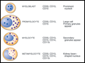 | Figure 60-2 Stages of neutrophil development shown schematically. G-CSF (granulocyte colony-stimulating factor) and GM-CSF (granulocyte-macrophage colony-stimulating factor) are critical to this process. Identifying cellular characteristics and specific cell-surface markers are listed for each maturational stage. |
view large |
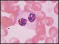
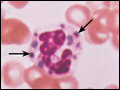
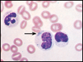 | Figure 60-3 Neutrophil band with Döhle body. The neutrophil with a sausage-shaped nucleus in the center of the field is a band form. Döhle bodies are discrete, blue-staining nongranular areas found in the periphery of the cytoplasm of the neutrophil in infections and other toxic states. They represent aggregates of rough endoplasmic... |
view large |
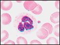 | Figure 60-4 Normal granulocyte. The normal granulocyte has a segmented nucleus with heavy, clumped chromatin; fine neutrophilic granules are dispersed throughout the cytoplasm. |
view large |
 | Figure 60-5 Pelger-Hüet anomaly. In this benign disorder, the majority of granulocytes are bilobed. The nucleus frequently has a spectacle-like, or "pince-nez," configuration. |
view large |
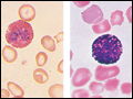 | Figure 60-6 Normal eosinophil and basophil. The eosinophil contains large, bright orange granules and usually a bilobed nucleus. The basophil contains large purple-black granules that fill the cell and obscure the nucleus. |
view large |
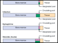 | Figure 60-7 Schematic neutrophil distribution and kinetics between the different anatomic and functional pools. |
view large |
 | Figure 60-8 Neutrophil travel through the pulmonary capillaries is dependent on neutrophil deformability. Neutrophil rigidity (e.g., caused by C5a) enhances pulmonary trapping and response to pulmonary pathogens in a way that is not so dependent on cell-surface receptors. Intraalveolar chemotactic factors, such as those caused by... |
view large |
 | Figure 60-9 Chédiak-Higashi syndrome. The granulocytes contain huge cytoplasmic granules formed from aggregation and fusion of azurophilic and specific granules. Large abnormal granules are found in other granule-containing cells throughout the body. |
view large |
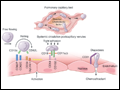
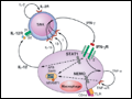 | Figure 60-10 Lymphocyte-macrophage interactions underlying resistance to mycobacteria and other intracellular parasites such as Salmonella. Mycobacteria infect macrophages, leading to the production of IL-12, which activates T or NK cells through its receptor, leading to production of IL-2 and... |
view large |