

PART 2: Cardinal Manifestations and Presentation of Diseases
SECTION 10 Hematologic Alterations
58 Bleeding and Thrombosis
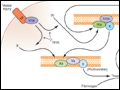 | Figure 58-1 Coagulation is initiated by tissue factor (TF) exposure, which, with factor (F)VIIa, activates FIX and FX, which in turn, with FVIII and FV as cofactors, respectively, results in thrombin formation and subsequent conversion of fibrinogen to fibrin. Thrombin activates FXI, FVIII, and FV, amplifying the coagulation... |
view large |
 | Figure 58-2 Fibrin formation and dissolution. (A). Fibrinogen is a trinodular structure consisting of 2 D domains and 1 E domain. Thrombin activation results in an ordered lateral assembly of protofibrils (B) with noncovalent associations. FXIIIa... |
view large |
 | Figure 58-3 Sites of action of the four major physiologic antithrombotic pathways: antithrombin (AT); protein C/S (PC/PS); tissue factor pathway inhibitor (TFPI); and the fibrinolytic system, consisting of plasminogen, plasminogen activator (PA), and plasmin. PT, prothrombin; Th, thrombin; FDP, fibrin(ogen) degradation products.... |
view large |
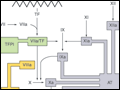
 | Figure 58-4 A schematic diagram of the fibrinolytic system. Tissue plasminogen activator (tPA) is released from endothelial cells, binds the fibrin clot, and activates plasminogen to plasmin. Excess fibrin is degraded by plasmin to distinct degradation products (FDPs). Any free plasmin is complexed with α2-antiplasmin... |
view large |
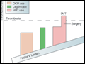 | Figure 58-5 Thrombotic risk over time. Shown schematically is an individual's thrombotic risk over time. An underlying factor V Leiden mutation provides a "theoretically" constant increased risk. The thrombotic risk increases with age and, intermittently, with oral contraceptive (OCP) or hormone replacement (HRT) use; other events... |
view large |
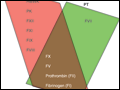 | Figure 58-6 Coagulation factor activity tested in the activated partial thromboplastin time (aPTT) in red and prothrombin time (PT) in green, or both. F, factor; HMWK, high-molecular-weight kininogen; PK, prekallikrein. |
view large |