

PART 2: Cardinal Manifestations and Presentation of Diseases
SECTION 9 Alterations in the Skin
52 Eczema, Psoriasis, Cutaneous Infections, Acne, and Other Common Skin Disorders
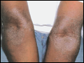 | Figure 52-1 Atopic dermatitis. Hyperpigmentation, lichenification, and scaling in the antecubital fossae are seen in this patient with atopic dermatitis. (Courtesy of Robert Swerlick, MD; with permission.) |
view large |
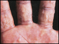 | Figure 52-2 Dyshidrotic eczema. This example is characterized by deep-seated vesicles and scaling on palms and lateral fingers, and the disease is often associated with an atopic diathesis. |
view large |
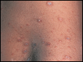
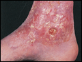 | Figure 52-3 Stasis dermatitis. An example of stasis dermatitis showing erythematous, scaly, and oozing patches over the lower leg. Several stasis ulcers are also seen in this patient. |
view large |
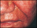 | Figure 52-4 Seborrheic dermatitis. Central facial erythema with overlying greasy, yellowish scale is seen in this patient. (Courtesy of Jean Bolognia, MD; with permission.) |
view large |
 | Figure 52-5 Lichen planus. An example of lichen planus showing multiple flat-topped, violaceous papules and plaques. Nail dystrophy as seen in this patient's thumbnail may also be a feature. (Courtesy of Robert Swerlick, MD; with permission.) |
view large |
 | Figure 52-6 Pityriasis rosea. In this patient with pityriasis rosea, multiple round to oval erythematous patches with fine central scale are distributed along the skin tension lines on the trunk. |
view large |
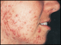 | Figure 52-7 Acne vulgaris. An example of acne vulgaris with inflammatory papules, pustules, and comedones. (Courtesy of Kalman Watsky, MD; with permission.) |
view large |
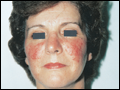 | Figure 52-8 Acne rosacea. Prominent facial erythema, telangiectasia, scattered papules, and small pustules are seen in this patient with acne rosacea. (Courtesy of Robert Swerlick, MD; with permission.) |
view large |