

PART 2: Cardinal Manifestations and Presentation of Diseases
SECTION 9 Alterations in the Skin
51 Approach to the Patient With a Skin Disorder
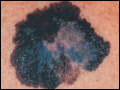 | Figure 51-1 Superficial spreading melanoma. This is the most common type of melanoma. Such lesions usually demonstrate asymmetry, border irregularity, color variegation (black, blue, brown, pink, and white), a diameter >6 mm, and a history of change (e.g., an increase in size or development of associated symptoms such as pruritus or... |
view large |
 | Figure 51-2 Nevomelanocytic nevus. Nevi are benign proliferations of nevomelanocytes characterized by regularly shaped hyperpigmented macules or papules of a uniform color. |
view large |
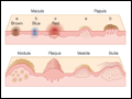 | Figure 51-3 A schematic representation of several common primary skin lesions (see |
view large |
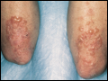
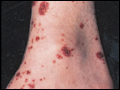 | Figure 51-4 Necrotizing vasculitis. Palpable purpuric papules on the lower legs are seen in this patient with cutaneous small vessel vasculitis. (Courtesy of Robert Swerlick, MD; with permission.) |
view large |
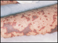 | Figure 51-5 Meningococcemia. An example of fulminant meningococcemia with extensive angular purpuric patches. (Courtesy of Stephen E. Gellis, MD; with permission.) |
view large |
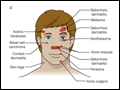 | Figure 51-6 A–D. The distribution of some common dermatologic diseases and lesions. |
view large |
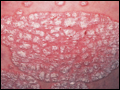 | Figure 51-7 Psoriasis. This papulosquamous skin disease is characterized by small and large erythematous papules and plaques with overlying adherent silvery scale. |
view large |
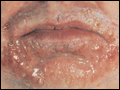
 | Figure 51-8 Dermatitis herpetiformis. This disorder typically displays pruritic, grouped papulovesicles on elbows, knees, buttocks, and posterior scalp. Vesicles are often excoriated due to associated pruritus. |
view large |
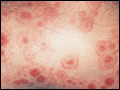 | Figure 51-9 Erythema multiforme. This eruption is characterized by multiple erythematous plaques with a target or iris morphology. It usually represents a hypersensitivity reaction to drugs (e.g., sulfonylamides) or infections (e.g., HSV). (Courtesy of the Yale Resident's Slide Collection; with... |
view large |
 | Figure 51-10 Allergic contact dermatitis (ACD). A. An example of ACD in its acute phase, with sharply demarcated, weeping, eczematous plaques in a perioral distribution. B. ACD in... |
view large |
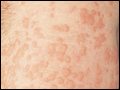 | Figure 51-11 Urticaria. Discrete and confluent, edematous, erythematous papules and plaques are characteristic of this whealing eruption. |
view large |
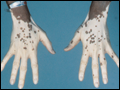 | Figure 51-12 Vitiligo. Characteristic lesions display an acral distribution and striking depigmentation as a result of loss of melanocytes. |
view large |