

PART 2: Cardinal Manifestations and Presentation of Diseases
SECTION 4 Disorders of Eyes, Ears, Nose, and Throat
29 Disorders of Smell and Taste
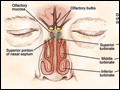 | Figure 29-1 Anatomy of the olfactory neural pathways, showing the distribution of olfactory receptors in the roof of the nasal cavity. [Copyright David Klemm, Faculty and Curriculum Support (FACS), Georgetown University Medical Center; used with permission.] |
view large |
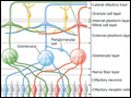 | Figure 29-2 Schematic of the layers and wiring of the olfactory bulb. Each receptor type (red, green, blue) projects to a common glomerulus. The neural activity within each glomerulus is modulated by periglomerular cells. The activity of the primary projection cells, the mitral and tufted cells, is modulated by granule cells,... |
view large |
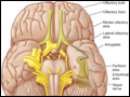 | Figure 29-3 Anatomy of the base of the brain showing the primary olfactory cortex. |
view large |
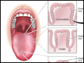 | Figure 29-4 Schematic of the taste bud and its opening (pore), as well as the location of buds on the three major types of papillae: fungiform (anterior), foliate (lateral), and circumvallate (posterior). TRC, taste receptor cell. |
view large |
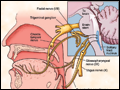 | Figure 29-5 Schematic of the cranial nerves that mediate taste function, including the chorda tympani nerve (CN VII), the glossopharyngeal nerve (CN IX), and the vagus nerve (CN X). [Copyright David Klemm, Faculty and Curriculum Support (FACS), Georgetown University Medical Center; used with... |
view large |
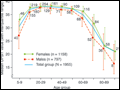 | Figure 29-6 Scores on the University of Pennsylvania Smell Identification Test (UPSIT) as a function of subject age and sex. Numbers by each data point indicate sample sizes. Note that women identify odorants better than men at all ages. (From Doty et al: Science 226:1421, 1984. Copyright 1984 American Association for the Advancement of... |
view large |