

PART 2: Cardinal Manifestations and Presentation of Diseases
SECTION 4 Disorders of Eyes, Ears, Nose, and Throat
28 Disorders of the Eye
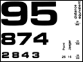 | Figure 28-1 The Rosenbaum card is a miniature, scale version of the Snellen chart for testing visual acuity at near. When the visual acuity is recorded, the Snellen distance equivalent should bear a notation indicating that vision was tested at near, not at 6 m (20 ft), or else the Jaeger number system should be used to report the... |
view large |
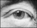 | Figure 28-2 Demonstration of a relative afferent pupil defect (Marcus Gunn pupil) in the left eye, done with the patient fixating on a distant target. A. With dim background lighting, the pupils are equal and relatively large. |
view large |
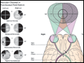 | Figure 28-3 Ventral view of the brain, correlating patterns of visual field loss with the sites of lesions in the visual pathway. The visual fields overlap partially, creating 120° of central binocular field flanked by a 40° monocular crescent on either side. The visual field maps in this figure were done with a... |
view large |
 | Figure 28-4 Retinal vasculitis, uveitis, and hemorrhage in a 32-year-old woman with Crohn's disease. Note that the veins are frosted with a white exudate. Visual acuity improved from 20/400 to 20/20 after treatment with intravenous methylprednisolone. |
view large |
 | Figure 28-5 Hollenhorst plaque lodged at the bifurcation of a retinal arteriole proves that a patient is shedding emboli from the carotid artery, great vessels, or heart. |
view large |
 | Figure 28-6 Central retinal artery occlusion combined with ischemic optic neuropathy in a 19-year-old woman with an elevated titer of anticardiolipin antibodies. Note the orange dot (rather than cherry red) corresponding to the fovea and the spared patch of retina just temporal to the optic disc. |
view large |
 | Figure 28-7 Hypertensive retinopathy with scattered flame (splinter) hemorrhages and cotton-wool spots (nerve fiber layer infarcts) in a patient with headache and a blood pressure of 234/120. |
view large |
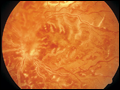
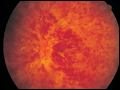 | Figure 28-8 Central retinal vein occlusion can produce massive retinal hemorrhage (“blood and thunder”), ischemia, and vision loss. |
view large |
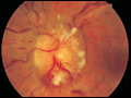
 | Figure 28-9 Anterior ischemic optic neuropathy from temporal arteritis in a 78-year-old woman with pallid disc swelling, hemorrhage, visual loss, myalgia, and an erythrocyte sedimentation rate of 86 mm/h. |
view large |
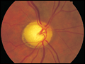
 | Figure 28-10 Retrobulbar optic neuritis is characterized by a normal fundus examination initially, hence the rubric “the doctor sees nothing, and the patient sees nothing.” Optic atrophy develops after severe or repeated attacks. |
view large |
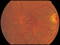
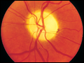 | Figure 28-11 Optic atrophy is not a specific diagnosis but refers to the combination of optic disc pallor, arteriolar narrowing, and nerve fiber layer destruction produced by a host of eye diseases, especially optic neuropathies. |
view large |
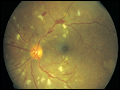
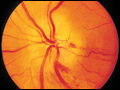
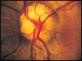
 | Figure 28-12 Papilledema means optic disc edema from raised intracranial pressure. This obese young woman with pseudotumor cerebri was misdiagnosed as a migraineur until fundus examination was performed, showing optic disc elevation, hemorrhages, and cotton-wool spots. |
view large |
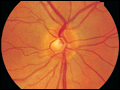
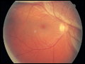
 | Figure 28-13 Optic disc drusen are calcified deposits of unknown etiology within the optic disc. They sometimes are confused with papilledema. |
view large |
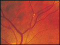
 | Figure 28-14 Retinal detachment appears as an elevated sheet of retinal tissue with folds. In this patient the fovea was spared, so acuity was normal, but a superior detachment produced an inferior scotoma. |
view large |
 | Figure 28-15 Glaucoma results in “cupping” as the neural rim is destroyed and the central cup becomes enlarged and excavated. The cup-to-disc ratio is about 0.7/1.0 in this patient. |
view large |
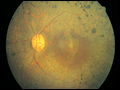
 | Figure 28-16 Age-related macular degeneration begins with the accumulation of drusen within the macula. They appear as scattered yellow subretinal deposits. |
view large |
 | Figure 28-17 Retinitis pigmentosa with black clumps of pigment in the retinal periphery known as “bone spicules.” There is also atrophy of the retinal pigment epithelium, making the vasculature of the choroid easily visible. |
view large |
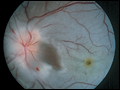
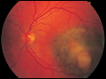 | Figure 28-18 Melanoma of the choroid, appearing as an elevated dark mass in the inferior temporal fundus, just encroaching upon the fovea. |
view large |
 | Figure 28-19 Left internuclear ophthalmoplegia (INO). A. In primary position of gaze the eyes appear normal. B. Horizontal gaze to the left is intact. |
view large |