

PART 2: Cardinal Manifestations and Presentation of Diseases
SECTION 3 Nervous System Dysfunction
22 Weakness and Paralysis
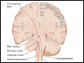 | Figure 22-1 The corticospinal and bulbospinal upper motor neuron pathways. Upper motor neurons have their cell bodies in layer V of the primary motor cortex (the precentral gyrus, or Brodmann's area 4) and in the premotor and supplemental motor cortex (area 6). The upper motor neurons in the primary motor cortex are... |
view large |
 | Figure 22-2 Lower motor neurons are divided into α and γ types. The larger α motor neurons are more numerous and innervate the extrafusal muscle fibers of the motor unit. Loss of α motor neurons or disruption of their axons produces lower motor neuron weakness. The smaller, less numerous γ motor... |
view large |
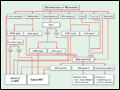 | Figure 22-3 An algorithm for the initial workup of a patient with weakness. CT, computed tomography; EMG, electromyography; LMN, lower motor neuron; MRI, magnetic resonance imaging; NCS, nerve conduction studies; UMN, upper motor neuron. |
view large |