

PART 2: Cardinal Manifestations and Presentation of Diseases
SECTION 1 Pain
15 Back and Neck Pain
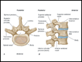 | Figure 15-1 Vertebral anatomy. (From A Gauthier Cornuelle, DH Gronefeld: Radiographic Anatomy Positioning. New York, McGraw-Hill, 1998; with permission.) |
view large |
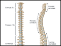 | Figure 15-2 Spinal column. (From A Gauthier Cornuelle, DH Gronefeld: Radiographic Anatomy Positioning. New York, McGraw-Hill, 1998; with permission.) |
view large |
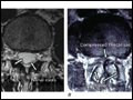
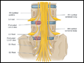 | Figure 15-3 Compression of L5 and S1 roots by herniated disks. (From Adams and Victor's Principles of Neurology, 9th ed. New York, McGraw-Hill, 2009; with permission.) |
view large |
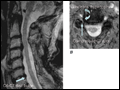
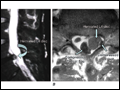 | Figure 15-4 Left L5 radiculopathy. A. Sagittal T2-weighted image on the left reveals disk herniation at the L4-5 level. B. Axial T1-weighted image shows paracentral disk herniation with displacement of the thecal sac medially and the left L5 nerve root posteriorly in the left lateral... |
view large |
 | Figure 15-5 Axial T2-weighted images of the lumbar spine. A. The image shows a normal thecal sac within the lumbar spinal canal. The thecal sac is bright. The lumbar roots are dark punctuate dots in the posterior thecal sac with the patient supine. |
view large |
 | Figure 15-6 Right C7 radiculopathy. A. Sagittal T2-weighted image shows mild disk bulging at C6-C7 and a mildly narrowed spinal canal, but no visible nerve root compression. B. Axial T2-weighted image. The combination of uncinate hypertrophy and facet... |
view large |